Chinese Journal of Clinical Pharmacology and Therapeutics ›› 2026, Vol. 31 ›› Issue (9): 1182-1192.doi: 10.12092/j.issn.1009-2501.2026.09.004
Junwei GAO1(
), Guijie MA2(
), Xiaomeng LIN1, Yingping DENG1, Chengqian YIN3, Chenguang WU3, Peng LIU4, Guanghui ZHONG1,*(
)
Received:2026-01-16
Revised:2026-04-03
Online:2026-09-26
Published:2026-10-08
Contact:
Guanghui ZHONG
E-mail:gaojwdr@163.com;maguijie@163.com;zgh20040712@126.com
CLC Number:
Junwei GAO, Guijie MA, Xiaomeng LIN, Yingping DENG, Chengqian YIN, Chenguang WU, Peng LIU, Guanghui ZHONG. Dioscin attenuates UUO-induced renal fibrosis by inhibiting the Akt/GSK-3β signaling pathway[J]. Chinese Journal of Clinical Pharmacology and Therapeutics, 2026, 31(9): 1182-1192.
| Species | Gene | Gene Primer sequence (5′-3′) | |
| Mouse | Fibronectin | F: AGGAACCGAGTACACCATCTA | R: GGGAAGCTCATCTGTCTTCTTC |
| α-SMA | F: GTCCCAGACATCAGGGAGTAA | R: TCGGATACTTCAGCGTCAGGA | |
| Vimentin | F: CTGCTAACTACCAGGACACTA | R: CAGGTTCAGGGAAGAAAA | |
| E-cadherin | F: CAGTTCCGAGGTCTACACCTT | R: TGAATCGGGAGTCTTCCGAAAA | |
| β-actin | F: ACCCTAAGGCCAACCGTGAAAAG | R: CATGAGGTAGTCTGTCAGGT | |
| Human | Fibronectin | F: CCACAGTGGAGTATGTGGTTAG | R: CAGTCCTTTAGGGCGATCAAT |
| α-SMA | F: CTATGAGGGCTATGCCTTG | R: GCTCAGCAGTAGTAACGAAGGA | |
| Vimentin | F: AGTCCACTGAGTACCGGAGAC | R: CATTTCACGCATCTGGCGTTC | |
| E-cadherin | F: CGAGAGCTACACGTTCACGG | R: GGGTGTCGAGGGAAAAATAGG | |
| β-actin | F: AGGCATCCTCACCCTGAAGTA | R: CACACGCAGCTCATTGTAGA | |
Table 1 Primer sequences for real-time PCR
| Species | Gene | Gene Primer sequence (5′-3′) | |
| Mouse | Fibronectin | F: AGGAACCGAGTACACCATCTA | R: GGGAAGCTCATCTGTCTTCTTC |
| α-SMA | F: GTCCCAGACATCAGGGAGTAA | R: TCGGATACTTCAGCGTCAGGA | |
| Vimentin | F: CTGCTAACTACCAGGACACTA | R: CAGGTTCAGGGAAGAAAA | |
| E-cadherin | F: CAGTTCCGAGGTCTACACCTT | R: TGAATCGGGAGTCTTCCGAAAA | |
| β-actin | F: ACCCTAAGGCCAACCGTGAAAAG | R: CATGAGGTAGTCTGTCAGGT | |
| Human | Fibronectin | F: CCACAGTGGAGTATGTGGTTAG | R: CAGTCCTTTAGGGCGATCAAT |
| α-SMA | F: CTATGAGGGCTATGCCTTG | R: GCTCAGCAGTAGTAACGAAGGA | |
| Vimentin | F: AGTCCACTGAGTACCGGAGAC | R: CATTTCACGCATCTGGCGTTC | |
| E-cadherin | F: CGAGAGCTACACGTTCACGG | R: GGGTGTCGAGGGAAAAATAGG | |
| β-actin | F: AGGCATCCTCACCCTGAAGTA | R: CACACGCAGCTCATTGTAGA | |
Fig.1 Dioscin attenuates renal pathological changes and collagen deposition in UUO mice A: representative photomicrographs of HE staining of kidney tissues from each group on day 7 after UUO operation (bar=50 μm). Representative photomicrographs of Masson's trichrome staining of kidney tissues from each group (bar=50 μm). Blue areas indicate collagen deposition. B: Left: scoring of renal tubular damage assessed by loss of the brush border, tubular dilation, inflammatory cell infiltration degree according to random HE-stained kidney cortical region sections. Right: scoring of Masson's trichrome staining assessed by percentage of blue-stained collagen area in each selected random cortical region field ($ \overline{x} $±s, n=5). cP<0.01, compared with UUO group.
Fig.2 Effect of Dioscin on the expression of EMT-related markers in kidneys of UUO mice A: representative images of immunohistochemical staining for α-SMA and FN in renal tissues (bar=50 μm). B: representative Western blot images showing the protein expression levels of α-SMA, Vimentin and E-cadherin in renal tissues. C: quantitative analysis of α-SMA, Vimentin and E-cadherin protein expression. D: mRNA relative expression levels of FN, α-SMA, and E-cadherin in renal tissues detected by RT-PCR ($ \overline{x} $±s, n=3). bP<0.05, cP<0.01, compared with UUO group.
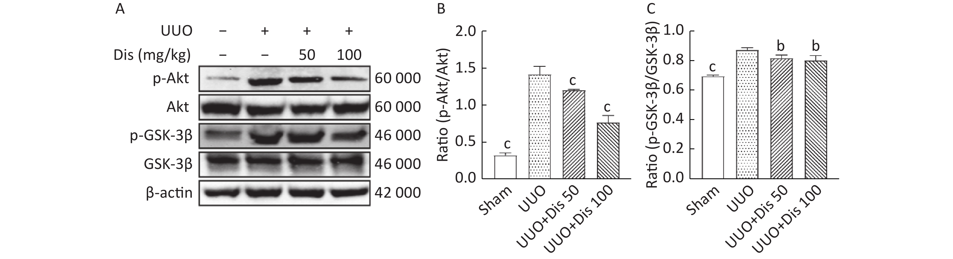
Fig.3 Effect of Dioscin on the expression of Akt/GSK-3β pathway proteins in kidneys of UUO mice A: representative Western blot images showing the protein expression levels of Akt, p-Akt (Ser473), GSK-3β, and p-GSK-3β (Ser9) in renal tissues. B: quantitative analysis of p-Akt, Akt, p-GSK-3β and GSK-3β protein expression and the ratios of p-Akt/Akt and p-GSK-3β/GSK-3β ($ \overline{x} $±s, n=3). bP<0.05, cP<0.01, compared with UUO group.
Fig.4 The Akt phosphorylation agonist SC79 aggravates TGF-β1-induced EMT-like phenotypic changes in HK-2 cells A: representative Western blot images showing the protein expression levels of α-SMA and Vimentin in HK-2 cells from each group. B: quantitative analysis of α-SMA and Vimentin protein expression. C: representative Western blot images showing the protein expression levels of Akt, p-Akt, GSK-3β, and p-GSK-3β. D: quantitative analysis of p-Akt, Akt, p-GSK-3β and GSK-3β protein expression and the ratios of p-Akt/Akt and p-GSK-3β/GSK-3β. E: mRNA relative expression levels of α-SMA, Vimentin, and E-cadherin detected by RT-PCR ($ \overline{x} $±s, n=3). bP<0.05, cP<0.01, compared with the TGF-β1 group.
Fig.5 Dis inhibits TGF-β1-induced EMT-like phenotypic changes and Akt/GSK-3β pathway activation in HK-2 cells A: cell viability of HK-2 cells treated with different concentrations of Dis for 24, 48, and 72 h, assessed by CCK-8 assay ($ \overline{x} $±s, n=8). cP<0.01, compared with the 0 μmol/L group. B: representative Western blot images showing the protein expression levels of α-SMA and Vimentin in HK-2 cells from different treatment groups. C: quantitative analysis of α-SMA and Vimentin protein expression. D: mRNA relative expression levels of α-SMA and Vimentin detected by RT-PCR. E: immunofluorescence staining for Vimentin (green) and E-cadherin (green). Scale bar=25 μm. F: representative Western blot images showing the protein expression levels of Akt, p-Akt, GSK-3β, and p-GSK-3β. G: quantitative analysis of the ratios of p-Akt/Akt and p-GSK-3β/GSK-3β ($ \overline{x} $±s, n=3). bP<0.05, cP<0.01, compared with the TGF-β1 group.
Fig.6 Dis counteracted the effects of SC79 on Akt/GSK-3β pathway activation and EMT marker expression in HK-2 cells A: representative Western blot images showing the protein expression levels of α-SMA and Vimentin. B: quantitative analysis of α-SMA and Vimentin protein expression. C: representative Western blot images showing the protein expression levels of Akt, p-Akt, GSK-3β, and p-GSK-3β. D: representative Western blot images showing the protein expression levels of Akt, p-Akt, GSK-3β, and p-GSK-3β ($ \overline{x} $±s, n=3). bP<0.05, cP<0.01, compared with the TGF-β1+SC79 group.
| 1 |
Law JP, Pickup L, Pavlovic D, et al. Hypertension and cardiomyopathy associated with chronic kidney disease: epidemiology, pathogenesis and treatment considerations[J]. J Hum Hypertens, 2023, 37 (1): 1- 19.
doi: 10.1038/s41371-022-00751-4 |
| 2 | Yan H, Xu J, Xu Z, et al. Defining therapeutic targets for renal fibrosis: exploiting the biology of pathogenesis[J]. Biomed Pharmacother, 2021, 143, 112115. |
| 3 | Zhang D, Zhang YH, Liu B, et al. Role of peroxisomes in the pathogenesis and therapy of renal fibrosis[J]. Metabolism, 2025, 166, 156173. |
| 4 | Hsieh YH, Tsai JP, Ting YH, et al. Rosmarinic acid ameliorates renal interstitial fibrosis by inhibiting the phosphorylated-AKT mediated epithelial-mesenchymal transition in vitro and in vivo[J]. Food Funct, 2022, 13 (8): 4641- 4652. |
| 5 |
Lu S, Chen X, Chen Y, et al. Downregulation of PDZK1 by TGF-β1 promotes renal fibrosis via inducing epithelial-mesenchymal transition of renal tubular cells[J]. Biochem Pharmacol, 2024, 220, 116015.
doi: 10.1016/j.bcp.2023.116015 |
| 6 | Seo JH, Lee HJ, Sim DY, et al. Honokiol inhibits epithelial-mesenchymal transition and hepatic fibrosis via activation of Ecadherin/GSK3β/JNK and inhibition of AKT/ERK/p38/β-catenin/TMPRSS4 signaling axis[J]. Phytother Res, 2023, 37 (9): 4092- 4101. |
| 7 | He S, Xia T, Guo Z, et al. Tracing the origin of myofibroblasts in kidney fibrosis[J]. Nat Commun, 2025, 17 (1): 653. |
| 8 | Li Y, Gao M, Yin LH, et al. Dioscin ameliorates methotrexate-induced liver and kidney damages via adjusting miRNA-145-5p-mediated oxidative stress[J]. Free Radic Biol Med, 2021, 169, 99- 109. |
| 9 | Zhang W, Lin L, Zhang Y, et al. Dioscin potentiates the antitumor effect of suicide gene therapy in melanoma by gap junction intercellular communication-mediated antigen cross-presentation[J]. Biomed Pharmacother, 2022, 150, 112973. |
| 10 | Wang Y, Yu D, Zhu S, et al. The genus Dioscorea L. (Dioscoreaceae), a review of traditional uses, phytochemistry, pharmacology, and toxicity[J]. J Ethnopharmacol, 2024, 329, 118069. |
| 11 | Dinesh Babu V, Suresh Kumar A, Sudhandiran G. Diosgenin inhibits TGF-β1/Smad signaling and regulates epithelial mesenchymal transition in experimental pulmonary fibrosis[J]. Drug Chem Toxicol, 2022, 45 (3): 1264- 1275. |
| 12 | Hosseinian S, Rad AK, Bideskan AE, et al. Thymoquinone ameliorates renal damage in unilateral ureteral obstruction in rats[J]. Pharmacological Rep, 2017, 69 (4): 648- 657. |
| 13 | Yin L, Li H, Liu Z, et al. PARK7 protects against chronic kidney injury and renal fibrosis by inducing SOD2 to reduce oxidative stress[J]. Front Immunol, 2021, 12, 690697. |
| 14 | 钟小冬, 杨军平. 肾间质纤维化的关键信号通路和血清学指标研究进展[J]. 光明中医, 2023, 38 (22): 4490- 4492. |
| 15 | Sun AB, Li FH, Zhu L, et al. TRPC6 knockout alleviates renal fibrosis through PI3K/AKT/GSK3B pathway[J]. Curr Med Sci, 2024, 44 (3): 589- 602. |
| 16 | Bao S, Chen T, Chen J, et al. Multi-omics analysis reveals the mechanism of action of ophiopogonin D against pulmonary fibrosis[J]. Phytomedicine, 2023, 121, 155078. |
| 17 | Lovisa S, Lebleu VS, Tampe B, et al. Epithelial-to-mesenchymal transition induces cell cycle arrest and parenchymal damage in renal fibrosis[J]. Nat Med, 2015, 21 (9): 998- 1009. |
| 18 | Minamida A, Nakata T, Kurose R, et al. Injured tubule derived CCN1 exacerbates renal congestion-mediated acute kidney injury and fibrosis[J]. Sci Rep, 2025, 15 (1): 20840. |
| 19 | Chen SL, Hu SW, Lin YY, et al. Boehmeria nivea extract (BNE-RRC) reverses epithelial-mesenchymal transition and inhibits anchorage-independent growth in tumor cells[J]. Int J Mol Sci, 2024, 25 (17): 9572. |
| 20 | Chiang CH, Lan TY, Hsieh JH, et al. Diosgenin reduces acute kidney injury and ameliorates the progression to chronic kidney disease by modifying the NOX4/p65 signaling pathways[J]. J Agric Food Chem, 2024, 72 (31): 17444- 17454. |
| 21 | Alhaj Sulaiman A, Katanaev VL. Beyond antioxidants: how redox pathways shape cellular signaling and disease outcomes[J]. Antioxidants, 2025, 14 (9): 1142. |
| 22 | Hong CE, Le D, Lee M, et al. Pro-apoptotic and anti-EMT activity of wild ginseng adventitious root extract in MDA-MB-231 TNBC cells: association with GSK-3β/β-catenin signaling[J]. Pharmaceuticals (Basel), 2026, 19 (2): 86. |
| 23 | Lee YJ, Han HJ. Troglitazone ameliorates high glucose-induced EMT and dysfunction of SGLTs through PI3K/Akt, GSK-3β, Snail1, and β-catenin in renal proximal tubule cells[J]. Am J Physiol Renal Physiol, 2010, 298 (5): F1263- F1275. |
| 24 | Kanlaya R, Peerapen P, Nilnumkhum A, et al. Epigallocatechin-3-gallate prevents TGF-β1-induced epithelial-mesenchymal transition and fibrotic changes of renal cells via GSK-3β/β-catenin/Snail1 and Nrf2 pathways[J]. J Nutr Biochem, 2020, 76, 108266. |
| 25 | Mao W, Yin H, Chen W, et al. Network pharmacology and experimental evidence reveal dioscin suppresses proliferation, invasion, and EMT via AKT/GSK3b/mTOR signaling in lung adenocarcinoma[J]. Drug Des Devel Ther, 2020, 14, 2135- 2147. |
| 26 | 李娟, 杨林. 尿毒清颗粒联合非布司他治疗高尿酸血症肾损害患者研究[J]. 实用医学杂志, 2024, 40 (10): 1418- 1422. |

| [1] | Tao WANG, Xiaowen CHENG, Zhirui ZHANG, Hao JIAO. Shikonin restrains TGF-β1/Smad signaling pathway to improve renal fibrosis [J]. Chinese Journal of Clinical Pharmacology and Therapeutics, 2026, 31(1): 48-54. |
| [2] | LIN Jingyi, HAN Rangyue, XU Linghui, TAN Ruizhi, SU Hongwei, WANG Li. Huangqi sanqi mixture inhibits lncRNA Gm51500/Adam12 axis to improve renal fibrosis in CKD [J]. Chinese Journal of Clinical Pharmacology and Therapeutics, 2025, 30(6): 750-762. |
| [3] | WU Xiaoxue, YE Yiping, LEI Zhendong, ZHANG Zunjing, CHEN Wenling. Icariin improves hypertensive renal fibrosis and injury through Cx32-Nox4 signaling pathway [J]. Chinese Journal of Clinical Pharmacology and Therapeutics, 2024, 29(8): 870-878. |
| [4] | LI Shixu, LI Linyun, WANG Xin, LI Ke, BIAN Hua. Effects of Ginkgo biloba extract on renal injury in rats with experimental renal failure through miR-145/FOXO1 axis [J]. Chinese Journal of Clinical Pharmacology and Therapeutics, 2023, 28(7): 728-735. |
| [5] | CHENG Xiaolong, HU Kunmei, HU Haoran,XU Hanghang, YANG Jieren, HAN Jun. Effects of Chinese medicine Yuxia capsule on renal fibrosis in spontaneously hypertensive rats [J]. Chinese Journal of Clinical Pharmacology and Therapeutics, 2018, 23(6): 632-639. |
| [6] | HU Kunmei, HU Haoran, SHEN Yuanyuan, LU Yining, YANG Jieren, HAN Jun. Effect of traditional Chinese medicine Yuxia capsule on renal fibrosis in spontaneously hypertensive rats and its relationship with TGF-β1/Smads signaling pathway [J]. Chinese Journal of Clinical Pharmacology and Therapeutics, 2017, 22(4): 387-393. |
| [7] | SU Jian, YIN Li-ping, ZHANG Xin, LI Bi-bo, LIU Li, LI Hui. Influence of rhein intervention on the expression of HGF and BMP7 in renal tissue of rats with chronical allograft nephropathy [J]. Chinese Journal of Clinical Pharmacology and Therapeutics, 2011, 16(10): 1114-1120. |
| [8] | WANG Jian-yun, YIN Xiao-xing. Relationship between transforming growth factor-β1 signal transduction and renal fibrosis of diabetes [J]. Chinese Journal of Clinical Pharmacology and Therapeutics, 2005, 10(4): 371-376. |
| [9] | WU Ji-Ning, LIU Bi-Cheng. Current researches of angiotensin receptor blockers in treatment of patients with chronic progressive renal disease [J]. Chinese Journal of Clinical Pharmacology and Therapeutics, 2004, 9(11): 1201-1204. |
| Viewed | ||||||
|
Full text |
|
|||||
|
Abstract |
|
|||||