Loading...
Welcome to Chinese Journal of Clinical Pharmacology and Therapeutics,Today is Chinese

Table of Content

    Volume 24 Issue 7
    26 July 2019
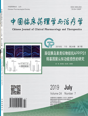
    Study on GIP analogues against the cognitive impairments in APP/PS1 transgenic mice
    YUAN Li, HAN Lingna, CHANG Yongli, ZHANG Cuiying
    2019, 24(7):  721-729.  doi:10.12092/j.issn.1009-2501.2019.07.001
    Asbtract ( 314 )   PDF (4366KB) ( 177 )  
    References | Related Articles | Metrics

    AIM: To investigate the neuroprotective effects of DAla2GIP-Glu-PAL on the cognitive impairment in APP/PS1 transgenic mice. METHODS: The nine-month-old male APP/PS1 mice and littermate WT mice were randomly divided into: WT-PBS, APP/PS1-PBS, WT-DAla2GIP-Glu-PAL and APP/PS1-DAla2GIP- Glu-PAL. APP/PS1 and WT mice were injected (i.p.) with PBS (0.01 mol/L) or DAla2GIP-Glu-PAL (5 μmol/L) at a volume of 0.2 mL once a day for 21 days prior to Morris experiments, then immunofluorescence staining, Western blot and ELISA experiments were performed.RESULTS:In Morris water maze test, the escape latency of APP/PS1 transgenic mice was significantly higher than that of WT mice from the 3rd to 5th day (P<0.01). In the probe trails experiment, compared with WT-PBS group mice, the swimming time in the target quadrant of APP/PS1-PBS was significantly decreased (P<0.01). Compared to the APP/PS1-PBS group, there were a significant decrease in the escape latency and an increase in swim time in target quadrant in the APP/PS1-DAla2GIP-Glu-PAL group. The fluorescence intensity of Aβ and GFAP in the hippocampus area of mice was detected by immunofluorescence staining, and the content of Aβ protein in mice was detected by Western blot. The value of APP/PS1-PBS group was significantly higher than that of WT-PBS group (P<0.01), and the value of APP/PBS-DAla2GIP-Glu-PAL group was significantly lower than that of APP/PS1-PBS group (P<0.01). In ELISA experiments, compared with the WT-PBS group, the content of IL-1β and TNF-α in the hippocampus area of the mice were significantly increased, and the content of insulin degrading enzyme was significantly decreased in APP/PS1-PBS group (P<0.01). Compared with the APP/PS1-PBS group, the content of IL-1β and TNF-α were significantly decreased. The content of insulin-degrading enzyme was significantly increased in APP/PBS-DAla2GIP-Glu-PAL group (P<0.01). CONCLUSION: DAla2GIP-Glu- PAL reverses cognitive function damage in APP/PS1 mice by inhibiting Aβ deposition, astrocyte proliferation and inflammatory factor secretion in hippocampus and up-regulating insulin-degrading enzyme content.

    Effects of autophagy on the protection of minocycline against cerebral ischemia-reperfusion injury in rats
    XIAO Shigeng, DONG Wenbin, YE Xiaodi, CHEN Aiying, CHENG Min, MIAO Yunping, ZHENG Gaoli
    2019, 24(7):  730-736.  doi:10.12092/j.issn.1009-2501.2019.07.002
    Asbtract ( 287 )   PDF (3155KB) ( 203 )  
    References | Related Articles | Metrics

    AIM: To investigate the effects of autophagy on the protection of minocycline against cerebral ischemia-reperfusion injury in rats. METHODS: Minocycline or Trimethyl adenine was injected to assess the neural function after the middle cerebral artery occlusion (MCAO) model established. The infarct volume was dertemined by TTC staining method at 24h after reperfusion. The ultrastructure of neurone and the quantity changes of autophagic vacuoles were observed by using transmission electron microscopy (TEM). The expression of protein LC3Ⅱ, Beclin1 and p62 were determined by Western blot. RESULTS:Minocycline (22.5 mg/kg, 45 mg/kg) significantly reduced rat nerve function scores and the cerebral infarction volume, while improved the expression of autophagy related proteins LC3Ⅱ and Beclin1 and increased the number of autophagosome and degradation of p62. Minocycline(90 mg/kg) could further increase the expression of LC3Ⅱ and Beclin1, with improvement of neural function and increase of infarct volume. Whereas the neuroprotective effects of minocycline(45 mg/kg) was reversed by the autophagy inhibitor 3-MA. CONCLUSION: Low dose minocycline attenuates cerebral ischemia reperfusion injury in rats, which might be mediated by up-regulating the protein LC3Ⅱand Beclin1 expression and promoting the degradation of p62/SQSTM l and then inducing autophagy; High dose minocycline aggravates cerebral ischemia reperfusion injury in rats by possible over activation of autophagy.

    Effects of curcumin on injury in hippocampal neurons and expression of blood corticosterone and SGK1 in hippocampal during global cerebral ischemia-reperfusion injury in hypertensive rats
    YU Chenchen, LIU Shuqun, LIU Jianlong, CAO Hong, LIU Xuejiao, CAI Xixi
    2019, 24(7):  737-743.  doi:10.12092/j.issn.1009-2501.2019.07.003
    Asbtract ( 303 )   PDF (3317KB) ( 202 )  
    References | Related Articles | Metrics

    AIM: To investigate the effects of curcumin on injury in hippocampal neurons and expression of blood corticosterone and serum-and glucocorticoid-inducible kinases1(SGK1) in hippocampal during global cerebral ischemia-reperfusion injury in hypertensive rats. METHODS: Male Wistar-Kyoto rats and male spontaneously hypertensive rats were randomLy divided into five groups (n=30): sham group (W-Sham and S-Sham), ischemia-reperfusion group (W-IR and S-IR), curcumin group (S-Cur). Rats were sacrificed at 3 h, 12 h, 1 d, 3 d, 7 d after reperfusion. Global brain ischemic model was established by 4-VO method. HE Staining was used to observe the vertebral cell morphology in hippocampal CA1 region. Nissl Stainning was applied to detect the average density of vertebral cell in hippocampal CA1 region. The corticosterone in blood and SGK1 in hippocampal were determined by ELISA. At 7d after reperfusion the behavior of the rats was observed. RESULTS:Compared with sham group , the ability to learn and remember was greatly decreased in ischemia-reperfusion group, the number of injured neurons was significantly enhanced , the corticosterone in blood was extremely enhanced(P<0.05), the expression of SGK1 in hippocampal was significantly increased(P<0.05). Compared with W-IR, the ability of S-IR to learn and remember was greatly decreased, the number of injured neurons was extremely enhanced, the corticosterone in blood was significantly increased(P<0.05), the expression of SGK1 in hippocampal was significantly decreased(P<0.05). The number of injured neurons was greatly declined in S-Cur, the ability to learn and remember was improved , the corticosterone in blood was extremely decreased(P<0.05), the expression of SGK1 in hippocampal was significantly increased(P<0.05). CONCLUSION: Inhibition of corticosterone in blood and overexpression of SGK1 in hippocampus may be involved in the mechanism by which curcumin reduces global cerebral IR injuty in hypertensive rats.

    Experimental study of Fuzheng Kangfu mixture combined with cisplatin on apoptosis of the gastric cancer cell SGC7901
    TENG Xiaojing, DONG Xueyan, WANG Zhiyi, YU Daojun, WANG Xianjun
    2019, 24(7):  744-749.  doi:10.12092/j.issn.1009-2501.2019.07.004
    Asbtract ( 293 )   PDF (3283KB) ( 189 )  
    References | Related Articles | Metrics

    AIM: To explore the effect of Fuzheng Kangfu mixture combined with cisplatin on promoting apoptosis and inhibiting proliferation of gastric cancer cell. METHODS: Twenty-eight nude mice were randomly divided into 4 groups as follows: the model control group, the group of Fuzheng, the cisplatin group, the group of cisplatin combined with Fuzheng. There were 7 nude mice in each group. The transplanted gastric cancer model of mice was established by subcutaneous injection of gastric cancer SCG7901 cell line. The Cisplatin as well as Fuzheng were administered drugs continuously for 7 days. The weight and tumor weight were measured and the tumor inhibition rate was calculated .The PCR and immunohistochemistry was applied to detect the apoptosis target of Bcl-2, Bax and Survivin as well as VEGF in nude mice. RESULTS:Compared with the model control group, the group of Fuzheng, the cisplatin group, the group of cisplatin combined with Fuzheng, the tumor weights of all the other nude mice were alleviated in different degrees (P<0.05). Among these, the tumor inhibition rate of the group of Cisplatin combined with Fuzheng was the highest (66.67%). In terms of cell inhibition, Fuzheng Kangfu mixture combined with cisplatin could significantly enhance the expression of Bax, and reduce the expressions of Bcl-2, Survivin, and vascular endothelial growth factor (VEGF) (P<0.05).CONCLUSION: Fuzheng Kangfu mixture combined with cisplatin has the effect of inhibiting tumor proliferation.

    Saponins from Rhizoma Panacis Majoris protect against cerebral ischemia and reperfusion injury through modulation of PI3K/Akt signaling pathway in mice
    DUAN Jinning, XIANG Changqing, QIAN Aihong, LI Jin, CHEN Xiuquan, HAN Yongfeng, HUANG Na, FAN Lili
    2019, 24(7):  750-758.  doi:10.12092/j.issn.1009-2501.2019.07.005
    Asbtract ( 269 )   PDF (4152KB) ( 176 )  
    References | Related Articles | Metrics

    AIM: To study the protective effect and possible mechanism of saponins from Rhizoma Panacis Majoris (SRPM) on cerebral ischemia-reperfusion (I/R) injury in mice. METHODS: Male Kunming mice were randomly divided into sham operation group, model group, SRPM (100 mg/kg), SRPM (200 mg/kg) and nimodipine group (2 mg/kg). The drug groups were given corresponding drugs by gavage, while the sham operation group and the model group were given 0.5% sodium carboxymethyl cellulose solution by gavage once a day for 7 consecutive days. After 7 days of administration, the middle cerebral artery occlusion model was established in mice in the sham operation group. After 2 hours of ischemia and 24 hours of reperfusion, the neurological deficit score, brain edema, infarct area and brain histomorphology were analyzed. The levels of ROS, LDH, SOD, GSH-Px, CAT and MDA in blood were detected. The protein levels of p-PI3K, PI3K, p-Akt, Akt Bcl-2, Bax, cytochrome C, cleaved-caspase-9 and cleaved-caspase-3 in brain tissue were detected by Western blot. RESULTS:SRPM (100 and 200 mg/kg) and nimodipine (2 mg/kg) could significantly improve neurological deficit score, brain edema, infarct area and brain histomorphology in cerebral I/R injury mice, reduce the levels of ROS, LDH and MDA, increase SOD, GSH-Px, CAT activities in serum, up-regulate the protein expressions of p-PI3K, p-Akt, Bcl-2, and Bcl-2/Bax ratio, down-regulate the protein expressions of Bax, cytochrome C, cleaved-caspase-9 and cleaved-caspase-3. CONCLUSION: The protective effect of SRPM on I/R injury mice may be related to its activation of PI3K/Akt pathway and inhibition of mitochondrial-mediated apoptotic pathway.

    Effects of propofol on spinal cord GRPR of intrathecal morphine-induced pruritus model in rats
    HENG Bingbing, DAI Shuyang, YANG Danfeng, BEEKOO Deepti, LIAN Qingquan, SHANGGUAN Wangning
    2019, 24(7):  759-765.  doi:10.12092/j.issn.1009-2501.2019.07.006
    Asbtract ( 290 )   PDF (3140KB) ( 174 )  
    References | Related Articles | Metrics

    AIM: To observe the effects of propofol on intrathecal morphine-induced pruritus in rats and explore the possible mechanism of propofol in treating intrathecal morphine-induced pruritus. METHODS: Thirty rats succeeded with intrathecal morphine-induced pruritus model, were randomly divided into 5 groups: 8 min group, 16 min group, 24 min group, 32 min group and 60 min group. 10 min after intrathecal injection with 40μg/kg morphine , rats were killed at 8 min, 16 min, 24 min, 32 min and 60 min thereafter, respectively to collect the dorsal spinal cord for Western blot. Forty-eightrats succeeded with intrathecal morphine-induced pruritus model, were randomLy divided into 4 groups: control group, propofol group, normal saline group and intralipid group. Same volume of normal saline 80 μL/kg, propofol 0.8 mg/kg, normal saline 80 μL/kg, intralipid 80 μL/kg were administered via the jugular vein to four groups respectively 10 min after intrathecal injection with normal saline or 40μg/kg morphine. Six rats were randomly selected from each group for pruritus behavior observation. The observation started from 30 min before up to 60 min after the intrathecal injection, and the injection scratching responses of the rat were recorded. The remaining rats in each group were executed after the intrathecal injection 42 min to collect the spinal cord for Western blot and immunohistochemistry to detect the protein concentration of GRPR. RESULTS:The peak of scratching frequency was at 10-20 min after morphine intrathecal injection, and almost disappeared at 60min. Compared with normal saline and intralipid groups, the scratching behavior was significantly decreased in propofol group (P<0.01); the expression of GRPR in spinal cord of control group and propofol group was lower (P<0.01). There was no significant difference of the expression of GRPR between normal saline group and intralipid group. CONCLUSION: Intrathecal injection of morphine-induced scratching behavior can be significantly alleviated by low-dose propofol. Propofol may significantly decrease the expression of GRPR in spinal cord in morphine-induced pruritus model.

    Lidocaine inhibits structural remodeling of vascular wall in diabetic rats by regulating inflammatory response
    SUN Aili, MA Yuetao, WANG Mingcang
    2019, 24(7):  766-772.  doi:10.12092/j.issn.1009-2501.2019.07.007
    Asbtract ( 302 )   PDF (2928KB) ( 150 )  
    References | Related Articles | Metrics

    AIM: To study the effect of lidocaine on the inflammatory response and vascular smooth muscle cells proliferation and migration of diabetic rats. METHODS: Serum, vascular tissue and primary cultured vascular smooth muscle cells of diabetic model rats and lidocaine-treated diabetic model rats were taken for study. RESULTS:Lidocaine could effectively inhibit the secretion of TNF-α, IL-8 and PAF inflammatory factors, it could be seen that lidocaine could reduce the inflammatory response induced by diabetic cardiovascular disease. Lidocaine could reduce the absorbance of Brdu kit, Ki-67 level, vimentin expression level and the migration area of smooth muscle cells in the scar experiment, indicating that lidocaine could effectively reduce the proliferation and migration of vascular smooth muscle cells. Lidocaine effectively inhibited STAT3 signaling pathway and reduced proliferation and migration of vascular smooth muscle cells. The results were statistically significant (P<0.05). CONCLUSION: Lidocaine can effectively reduce the inflammatory response in diabetic rat model by inhibiting the activation of AKT-STAT3 signaling pathway, and at the same time reduce the abnormal proliferation and migration of vascular smooth muscle cells, thereby improving the vascular wall structure reconstruction. Lidocaine may be a potential therapeutic agent for diabetic cardiovascular disease induced by inflammatory response.

    Inhibitory effect of vemurafenib on UGT1A1-mediated irinotecan metabolism
    WEN Chunjie, ZHANG Ying, WU Lanxiang, ZHOU Honghao
    2019, 24(7):  773-777.  doi:10.12092/j.issn.1009-2501.2019.07.008
    Asbtract ( 307 )   PDF (1903KB) ( 183 )  
    References | Related Articles | Metrics

    AIM: To study the inhibitory effect of vemurafenib on UDP-glucuronosyltransferases 1A1 (UGT1A1)-mediated irinotecan metabolism, and predict the risk of drug-drug interactions (DDI) by in vitro-in vivo extrapolation (IV-IVE). METHODS: A panel of human liver microsomes (HLMs) and recombinant human UGT1A1 were used to characterize the inhibitory effect of vemurafenib on human UGT1A1-mediated glucuronidation of SN-38, the active metabolite of irinotecan. The half maximum inhibitory concentration (IC50) and the constant of inhibition kinetics (Ki) were obtained, and the potential risk of DDI was predicted based on in vitro parameters. RESULTS:Vemurafenib had strong non-competitive inhibitory effect on UGT1A1, the IC50 value was 4.35 μmol/L, and the inhibition kinetic constant Ki value was 9.77 μmol/L. The area under the curve (AUC) ratio of SN-38 can be increased by 7% to 149% at the oral dose of 960 mg twice daily. CONCLUSION: The strong inhibition of vemurafenib on UGT1A1 leads to reduction of the UGT1A1-mediated irinotecan metabolism, and increases the risk of DDI.

    Correlation analysis of common bacterial resistance and antimicrobial use in a tertiary hospital
    SHENG Xuehe,XU Jinying,WANG Youlin,YIN Qin
    2019, 24(7):  778-785.  doi:10.12092/j.issn.1009-2501.2019.07.009
    Asbtract ( 544 )   PDF (3117KB) ( 206 )  
    References | Related Articles | Metrics

    AIM: To investigate the correlation between antimicrobial drug use and bacterial drug resistance, and to provide reference basis for clinical rational use of antimicrobial drugs. METHODS: The usage of commonly used antibacterial drugs and the drug resistance rate of bacteria isolated from clinical specimens in the hospital from 2012 to 2017 were investigated and statistically analyzed using SPSS 20.0 software.RESULTS:Among the common gram-negative bacteria in the hospital, the drug resistance rate of escherichia coli was correlated with DDDs of amikacin and imipenem (r=-0.957,P=0.011) and r=0.881 (P=0.048), respectively. The drug resistance rate of klebsiella pneumoniae was correlated with DDDs of imipenem (r=0.949,P=0.014).Among gram-positive bacteria, the drug resistance rate of staphylococcus haemolyticus was correlated with DDDs of levofloxacin, and the correlation coefficient was r=0.975 (P=0.025).CONCLUSION: There is a correlation between the amount of antimicrobial drugs and bacterial drug resistance.

    Effects of sulforaphane-induced Akt/p70S6K signal transduction pathway on apoptosis of human nasopharyngeal cancer cells CNE-2
    ZHAO Li, HUA Xia, TAN Ye, HU Chenglian
    2019, 24(7):  786-791.  doi:10.12092/j.issn.1009-2501.2019.07.010
    Asbtract ( 276 )   PDF (2459KB) ( 205 )  
    References | Related Articles | Metrics

    AIM: To study the effect of sulforaphane on the proliferation and apoptosis of CNE-2 cells, and to investigate the role of sulforaphane in the Akt/p70S6K signaling pathway.  METHODS: CCK-8 test was used to detect cellular growth of CNE-2. Flow cytometry was carried out to observe apoptosis and cell cycle status. Expression levels of several cell cycle associated proteins and Akt/p70S6K pathway related proteins were determined by Western blot.RESULTS:Cell proliferation inhibitory rates and apoptotic rates of CNE-2 cells in various doses of sulforaphane groups were significantly increased as compared with control group with a dose-dependent manner (P<0.05). Flow cytometry results showed that sulforaphane arrestted NPC cells at the S-phases and G2/M-phases, and the expression levels of Cyclin A, Cyclin B, Cyclin D, Cyclin E and CDK/p34 were greatly decreased in sulforaphane group as compared with control group (P<0.05). Moreover, the expression levels of p-Akt and p70S6K in sulforaphane group were also decreased as compared with control group (P<0.05). However, the expression levels of these proteins above in sulforaphane+ IGF-I group unchanged as compared with control group (P>0.05). CONCLUSION: Sulforaphane exerts anti-proliferative and pro-apoptotic effects on CNE-2 cells through interfering with the Akt/p70S6K signaling pathways.

    Efficiency of GnRH agonist trigger on embryo cultural outcomes in IVF subjects with diminished ovarian reserve
    ZHANG Ling, XU Weihai, HUANG Qiongxiao, FU Xiaohua, ZHANG Lin, LI Shishi, ZHU Jing, WU Ruifang, SHU Jing
    2019, 24(7):  799-804.  doi:10.12092/j.issn.1009-2501.2019.07.012
    Asbtract ( 299 )   PDF (2501KB) ( 191 )  
    References | Related Articles | Metrics

    AIM: To compare the effect of two trigger protocols-GnRH agonist and hCG on in vitro fertilization cultural outcome in patients with diminished ovarian reserve (DOR). METHODS: The patients with DOR undertaking the first cycle of IVF treatment in our IVF center were recruited for this study, and those with GnRH antagonist down-regulation stimulation excluded. All subjects were analyzed in two groups-GnRHa and hCG. The outcomes of oocyte retrieval, normal fertilization, embryo development were compared between the two groups. RESULTS:Comparison of GnRHa and hCG between the two groups showed no significant difference in age, BMI, index of basal function of ovary and the related parameters of treatment. In group GnRHa, the normal fertilization rate (% egg number), available embryo rate (% egg number), good embryo rate (% egg number) and good embryo per embryo were 73.03%, 63.10% and 42.24% respectively, which were significantly higher than those of the hCG. However, the aforementioned significance did not existed after applying the model of generalized estimation equation for the balancing age, ovarian reserve function, infertility factors and treatment regimen. CONCLUSION:GnRHa trigger may result in an equal laboratory culture outcomes to hCG, which need to be further confirmed by prospective studies.

    Effects of weekly liposome paclitaxel combined with tiggio on serum tumor markers and soluble e-cadherin in elderly patients with advanced gastric cancer
    DONG Jinliang
    2019, 24(7):  805-809.  doi:10.12092/j.issn.1009-2501.2019.07.013
    Asbtract ( 386 )   PDF (2045KB) ( 171 )  
    References | Related Articles | Metrics

    AIM: To analyze the effect of weekly liposome paclitaxel combined with tiggio on serum tumor markers and soluble e-cadherin (sEC) in elderly patients with advanced gastric cancer. METHODS: Eighty-seven elderly patients with advanced gastric cancer were divided into 41 control groups and 46 treatment groups according to the random number table. In the control group, olififloxacin was intravenously instilled at 130 mg/m2 each time, the first day of each cycle was 21 days, and the course of treatment was continued for 3 courses; tiggio each time 50 mg (body surface area>1.25 m-2-1.5 m-2) or 60 mg (body surface area≥1.5 m-2), oral, 1-28 d per cycle, 6 weeks for 1 course, continuous treatment for 2 courses. The usage of tiggio in the treatment group was the same as that in the control group; the liposome paclitaxel was intravenously infused at 60 mg/m2 each time, at the first, eighth, fifteen, and twenty-second days of the cycle, and one course of treatment was 6 weeks. Then clinical efficacy, serum levels of tumor markers, sEC before and after treatment, adverse reactions and survival in both group were compared.RESULTS:After treatment, the disease control rates in the treatment group and the control group were 65.22% (30/46) and 56.10% (23/46), 1-year survival rate was 58.69% (27/46) and 48.78% (20/41) , there was no statistical difference (P>0.05). Serum levels of carcinoembryonic antigen (CEA) in the treatment group and the control group were (9.37±2.03) and (8.99±1.48) ng/mL, carbohydrate antigen 125 (CA125) was (24.08±3.98) and (23.22±3.86) U/mL, the glucose chain antigen 19-9 (CA19-9) was (14.57±2.81) and (15.30±2.22) U/mL, sEC was (1 903.87±250.17) and (1 934.37±199.46) ng/mL, there was no statistical difference (P>0.05). The adverse reactions in the both groups were mainly nausea and vomiting, anemia, thrombocytopenia, granulocytopenia, diarrhea and liver and kidney dysfunction, adverse reaction rate in treatment group and the control group were statistically significant differences (P<0.05). CONCLUSION:The effect of weekly liposome paclitaxel combined with tiggio on elderly patients with advanced gastric cancer is confirmed, it can reduce the level of serum tumor markers and sEC, and patient tolerance is better.

    Clinical efficacy of sacubitril valsartan in the treatment of chronic heart failure
    FAN Jianfeng,FANG Yi,ZHENG Chunhua
    2019, 24(7):  810-814.  doi:10.12092/j.issn.1009-2501.2019.07.014
    Asbtract ( 546 )   PDF (2195KB) ( 206 )  
    References | Related Articles | Metrics

    AIM: To investigate the clinical effects of sacubitril valsartan and benazepril hydrochloride on heart function in patients with heart failure reduced ejection fraction(HFrEF)after two months of treatment. METHODS: Patients (52 cases) with heart failure reduced ejection fraction hospitalized in Third Affiliated Hospital of Nanchang University from November 2017 to September 2018 were divided into control (25 cases) and treatment (27cases) groups according to different treatments. Two groups of patients were given conventional anti-heart failure treatment. The control group was treated with benazepril hydrochloride 10 mg orally once a day and the dose was doubled every 2 weeks until the target dose was 20 mg orally 1time per day. The treatment group was treated with sacubitril valsartan 50 mg orally twice daily after stop ACEI 36 hours, and the dose was doubled every 2 weeks until the target dose was 200 mg orally 2 times per day. Patients in two groups were treated for 4 weeks. After treatment, the clinical efficacy was evaluated, and 6 min walking distance, the NT-pro BNP levels, LVEF,LVEDD,MLHFQ scores in two groups before and after treatment were compared.RESULTS:After treatment, the clinical efficacy in the control group was 72.00%, which was significantly lower than 92.60% in the treatment group (P<0.05). After treatment, the 6 min walking distance in two groups were significantly increased.The NT-pro BNP levels, LVEF,LVEDD,and MLHFQ scores were decreased, and the difference was statistically significant in the same group (P<0.05). The treatment group was significantly improved compared with control group (P<0.05). CONCLUSION:Sacubitril valsartan has significant curative effect and high safety in the treatment of chronic heart failure.

    Effects of dexmedetomidine on cognitive dysfunction and attentional network function in patients with ischemic cerebrovascular disease
    ZHANG Rui
    2019, 24(7):  815-820.  doi:10.12092/j.issn.1009-2501.2019.07.015
    Asbtract ( 300 )   PDF (2567KB) ( 178 )  
    References | Related Articles | Metrics

    AIM: To observe the effects of dexmedetomidine on postoperative cognitive dysfunction and attention network function in patients with ischemic cerebrovascular disease.  METHODS: A total of eighty-six patients with ischemic cerebrovascular disease who were admitted to our hospital from September 2015 to February 2018 were randomly divided into observation group and control group, with 43 cases in each group. In the observation group, dexmedetomidine was pumped intravenously before anesthesia, and the same dose of saline was pumped into the control group. The heart rate (HR), systolic blood pressure (SBP) and diastolic blood pressure (DBP) before operation (T0), after cannulation (T1), 30 min after operation(T2), end of surgery(T3), the cognitive function before operation, 6 h after operation, 24 h after operation, 72 h after operation, and 1 week after operation (assessed using MMSE scale), incidence of POCD, the attention network function (including alerting network efficiency, orienting network efficiency, executive control network efficiency, average response time, and accuracy) before operation and 72 h after operation were compared between the two groups. RESULTS:The HR, SBP and DBP at T1 and T2 in both groups were significantly lower than before surgery (P<0.05), and returned to the preoperative equivalent level at T3 (P>0.05). The HR at T1 and T2 in the observation group was significantly lower than that in the control group, and the SBP and DBP were significantly higher than those in the control group (P<0.05). The cumulative incidence of POCD in the observation group within 1 week after surgery was 9.30%, which was significantly lower than 27.91% in the control group (P<0.05). The MMSE scores of the observation group at 6 hours and 24 hours after operation were significantly lower than that before operation(P<0.05), and recovered to the preoperative level at 72 hours after operation(P>0.05). The MMSE scores of the control group at 6 hours, 24 hours, and 72 hours after operation were significantly lower than that before the operation (P<0.05). The MMSE score at one week after operation returned to the preoperative level (P>0.05). The alerting network efficiency, orienting network efficiency, and correct rate at 3 d after surgery of the two groups were significantly reduced than 1 d before surgery (P<0.05), the executive control network efficiency, and the average response time were significantly higher than 1 d before surgery (P<0.05). The alerting network efficiency, orienting network efficiency and accuracy of the observation group at 3 d after operation were significantly higher than those of the control group, and the executive control network efficiency and the average response time were significantly lower than those of the control group (P<0.05). CONCLUSION:The dexmedetomidine can slow down heart rate and reduce hemodynamic fluctuations, improve postoperative cognitive function and attention network function in patients with ischemic cerebrovascular disease.

    Review and economic analysis of preoperative antimicrobial application during the period of type I incision operation in a cancer hospital
    YANG Jingmo, ZHANG Bo, SUN Yancai
    2019, 24(7):  821-825.  doi:10.12092/j.issn.1009-2501.2019.07.016
    Asbtract ( 285 )   PDF (1947KB) ( 176 )  
    References | Related Articles | Metrics

    AIM: To analyze the surgical antimicrobial prophylaxis and to evaluate the effectiveness of different antimicrobial solutions about type I incision operation in a cancer hospital. METHODS: Three-hundred cases of clean surgery in our hospital from January to December in 2017 were involved and the data of the surgical antimicrobial prophylaxis were retrospective collected and evaluated.The cost-effectiveness analysis method was applied to compare the ratio of cost to effectiveness between the three solutions. RESULTS:The utilization ratio of antibiotics in clean surgery was 16.33%, which met the hospital requirements. Cefazolin (55.11%) and Cefuroxime (28.57%) were mainly preventive use in the 49 cases of antibiotics. DUI values of antibiotics for prophylaxis were ≤1, antibiotics dosage for single day was reasonable. The inappropriate use of antibiotics was mainly manifested in “unsuitable varieties”, which accounted for 63.64% of the total irrational rate. CONCLUSION: From the perspective of ecnnomic medication, the preoperative uses of cefazolin in patients treated with clean surgery was a better option.

    Research progresses of wound repair by exosomes derived from mesenchymal stem cells
    LIU Qingwu, CHEN Jia, MENG Yujiao, FENG Fang, GUO Xiwei, GUO Jianning, ZHANG Jinchao, LIN Yan, HE Xiujuan, LI Ping
    2019, 24(7):  826-832.  doi:10.12092/j.issn.1009-2501.2019.07.017
    Asbtract ( 375 )   PDF (3339KB) ( 186 )  
    References | Related Articles | Metrics

    Exosomes are membranous vesicles secreted by cells and promote wound healing by paracrine after secretion from stem cells. This article reviews recent advances of wound repair by exsosomes derived from mesenchymal stem cells. By regulating inflammation, promoting fibroblast proliferation and angiogenesis, regulating extracellular matrix remodeling, and inhibiting scar formation, mesenchymal stem cell exosomes can promote wound repair. Furthermore, engineered stem cells can increase their exosomes efficacy and yield, and provide great advantages to the clinical application of exosomes.

    Advances in extraction and mechanism of antitumor active components from Fagopyrum dibotrys
    LI Hongli, WEN Dandan, ZHOU Meiliang, WANG Haihua, PENG Xixu, GAO Lichen
    2019, 24(7):  833-840.  doi:10.12092/j.issn.1009-2501.2019.07.018
    Asbtract ( 325 )   PDF (3230KB) ( 194 )  
    References | Related Articles | Metrics

    Fagopyrum dibotrys is a medicinal plant of Buckwheat in Polygonaceae collected in Pharmacopoeia. It is widely distributed in the central and southwest of China. The extraction process of its antitumor active ingredients by immersion method is lagging behind. The main antitumor active ingredients, flavonoids and phenols, are concentrated in the rhizome. The main antitumor mechanisms of buckwheat are inhibiting the proliferation and migration of tumor cells, besides, inducing apoptosis and autophagy of cancer cells. In this paper, we reviewed the research progress on the antitumor chemical components, the extraction of active ingredients and the antitumor mechanisms of Fagopyrum dibotrys in order to provide references for further research on antitumor active ingredients and antitumor effects of Fagopyrum dibotrys.